The MethylFlash Methylated DNA Quantification Equipment (Colorimetric) is an entire set of optimized buffers and reagents to colorimetrically quantify international DNA methylation by particularly measuring ranges of 5-methylcytosine (5-mC) in an ELISA-like microplate-based format. In comparison with LUMA or LINE-1, Alu, and LTR-based assays, MethylFlash™ know-how instantly quantifies precise international DNA methylation.
This package can be particularly optimized for paired use with the MethylFlash Hydroxymethylated DNA Quantification Equipment (Colorimetric) for concurrently quantifying each methylated DNA and hydroxymethylated DNA.
Product Options
The MethylFlash™ Methylated DNA Quantification Equipment (Colorimetric) is an additional refinement of our earlier Methylamp™ international DNA methylation quantification kits by simplifying the workflow and enhancing the consistency of outcomes. It makes use of an revolutionary methodology to allow background indicators to be extraordinarily low, eliminating the plate drying and blocking steps. In comparison with chromatography-based strategies comparable to HPLC or mass spectrometry, this product supplies a cheap strategy to precisely measure ranges of 5-methylcytosine. The package has the next benefits and options:
Colorimetric assay with easy-to-follow steps for comfort and velocity. The complete process might be completed inside four hours.
Revolutionary package composition allows background indicators to be extraordinarily low, which eliminates the necessity for plate blocking and permits the assay to be easy, correct, dependable, and constant.
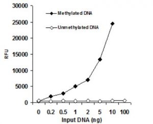
Excessive sensitivity, of which the detection restrict might be as little as 0.2 ng of methylated DNA and accepts as little as 50 ng of enter genomic DNA.
Optimized antibody and enhancer options enable excessive specificity to 5mC, with no cross-reactivity to unmethylated cytosine and no or negligible cross-reactivity to hydroxymethylcytosine throughout the indicated focus vary of the pattern DNA.
Common optimistic and adverse controls are included, that are appropriate for quantifying methylated DNA from any species.
Strip-well microplate format makes the assay versatile: handbook or excessive throughput evaluation.
Precept & Process
The MethylFlash Methylated DNA Quantification Equipment (Colorimetric) comprises all reagents crucial for the quantification of world DNA methylation. On this assay, DNA is sure to strip wells which can be particularly handled to have a excessive DNA affinity.
The methylated fraction of DNA is detected utilizing seize and detection antibodies after which quantified by an ELISA-like response by studying the absorbance in a microplate spectrophotometer at 450 nm. The quantity of methylated DNA is proportional to the OD depth measured, which might be calculated with the package’s included formulation for relative methylation standing of two totally different DNA samples or absolute quantification of 5-methylcytosine (5mC) utilizing an ordinary curve.
methylflash methylated dna quantification package
Simple, Quick, and Versatile
The complete colorimetric assay has easy-to-follow steps for comfort and velocity, which might be accomplished in four hours. The strip-well microplate format permits for a versatile assay in handbook or excessive throughput evaluation. Common optimistic and adverse controls are included with the package for quantifying methylated DNA from any species comparable to mammals, vegetation, fungi, micro organism, and viruses.
Secure and Handy
All of the wanted reagents, together with adverse controls and optimistic controls, for quantification of world DNA methylation are conveniently packaged within the package. The direct colorimetric quantification of DNA samples replaces out of date or inferior strategies and eliminates the necessity for DNA digestion/denaturation, radioactivity, extraction, or chromatography.
Extremely Delicate and Particular
The novel process and proprietary package compositions enable for correct quantification of methylated DNA to be achieved with excessive sensitivity and specificity. The detection restrict of the enter DNA might be as little as 0.2 ng of methylated DNA.
Comparability of Out there ELISA-based World DNA Methylation Assay Strategies
MethylFlash(Colorimetric)
Competitor Z
Competitor C
Assay Precept
Direct ELISA
Direct ELISA
Oblique ELISA
Format
96-well plate
96-well plate
96-well plate
Sensitivity
Glorious detection restrict: 0.2 ng of 5-mC DNA
Good detection restrict: >0.5 ng of 5-mC DNA
Very poor detection restrict: >1000 ng of 5-mC DNA
S/N Ratio
>20 with very low background
<four with excessive background
N/A*
Procedural Comfort
No want for plate blocking and denaturation of DNA
Requires plate blocking and DNA denaturation
Requires plate blocking, DNA digestion, and antibody aggressive incubation in an extra plate
Protocol Time
<four hr
<four hr
>eight hr
DNA Sort & Species
Common for any species, each ssDNA and dsDNA
Human and mouse ssDNA solely
Numerous species
Specificity
Particular to 5-mC solely
Cross-reaction to 5-hmC
N/A*
Quantitation Sort
Each relative and absolute quantification
Relative
Relative
Commonplace Management
Secure, quantitative in absolute quantity of 5-mC, and might be universally used for any species
Unstable, can’t be quantitative in quantity, and may solely be used for human or mouse
N/A*
Minimal Enter Quantity
50 ng
100 ng
1000 ng
Accuracy of Detection
Excessive, steady and quantified commonplace management. Confirmed shut correlation with LC-MS evaluation by customers
Unclear, unstable and un-quantified commonplace management
N/A*
% 5-mC calculation
Easy
Sophisticated
Not Offered
Patented Methodology
Sure
No
No
Recognition
Very excessive revealed quotation rely
Low revealed quotation rely
Low revealed quotation rely
Help Experience
Since 2006
Since 2013
Since 2011
DNA Methylation Quantification
DNA methylation performs an essential position in regular organismal improvement and in mobile differentiation in greater organisms. Gene expression, in addition to the event of practically all kinds of most cancers, are additionally tied to DNA methylation.
For instance, international lower in 5-methylcytosine content material (DNA hypomethylation) is probably going attributable to methyl-deficiency as a consequence of quite a lot of environmental influences and has been proposed as a molecular marker in a number of organic processes comparable to most cancers. World quantification of DNA methylation is essential for understanding the roles that gene expression and silencing play within the improvement of most cancers and different ailments.
Description: CCL11 is a potent eosinophil chemoattractant that was originally purified from bronchoalveolar lavage fluid of guinea pigs sensitized by aerosol challenge with ovalbumin. Mouse CCL11 cDNA encodes a 97 amino acid (aa) precursor protein from which the aminoterminal 23 aa are cleaved to generate the 74 aa mature mouse CCL11. At the protein sequence level, mature mouse CCL11 is approximately 60% identical to mature human and guinea pig CCL11. In addition, mouse CCL11 also shows high aa sequence identity to members of the MCP family. Mouse CCL11 is chemotactic for eosinophils, but not mononuclear cells or neutrophils. CCL11 mRNA is expressed in a variety of tissues. The expression of CCL11 mRNA is induced in cultured endothelial cells in response to IFNγ. In addition, CCL11 mRNA is also induced in response to the transplantation of IL4secreting tumor cells. The CC chemokine receptor 3 (CCR3) has been identified as a specific human CCL11 receptor.
Description: CCL12 is a cloned mouse CC chemokine most closely related to human MCP1 (66% amino acid (aa) sequence identity in the mature protein). Mouse CCL12 encodes a 104 aa residue precursor protein with a 22 aa residue predicted hydrophobic signal sequence that is cleaved to generate a 82 aa residue mature protein. CCL12 is expressed constitutively in the thymus and lymph nodes. Under inflammatory conditions, CCL12 expression is also induced in activated macrophages and mast cells. The gene for mouse MCP1 has been mapped to the CC chemokine cluster on chromosome 11. Recombinant CCL12 has been shown to be a potent chemoattractant for monocytes and lymphocytes but not neutrophils. At high concentrations, CCL12 will also chemoattract eosinophils. CCL12 has been found to be a functional ligand for CCR2 but not CCR1, CCR3, or CCR5.
Description: Human thymus and activationregulated chemokine (TARC) also known as CCL17, is a novel CC chemokine identified using a signal sequence trap method. Mouse CCL17 was discovered as a dendritic cell (DC) specific gene by differentiation RNA display. Mouse CCL17 cDNA encodes a highly basic 93 amino acid (aa) residue precursor protein with a 23 aa residue putative signal peptide that is cleaved to generate the 70 aa residue mature secreted protein. Among CC chemokine family members, CCL17 has approximately 24-29% amino acid sequence identity with RANTES, MIP1α, MIP1β, MCP1, MCP2, MCP3, and I309. The gene for human CCL17 has been mapped to chromosome 16q13 rather than chromosome 17 where the genes for many human CC chemokines are clustered. Mouse CCL17 is constitutively expressed in thymic DC, and at a lower level in lymph node DC in the lung. Recombinant CCL17 has been shown to be chemotactic for T cell lines and antigen-primed T helper cells. In humans, CCL17 was identified to be a specific functional ligand for CCR4 and CCR8, receptors that are selectively expressed on T cells.
Description: CCL19/MIP3β, also known as ELC (EBI1Ligand Chemokine), is a β chemokine that binds specifically to the chemokine receptor CCR7/EBI1/ BLR2. Mouse (human) CCL19 cDNA encodes a 108 (98) amino acid precursor protein with a predicted 25 (21) aa signal peptide that is cleaved to form the 83 (77) aa mature secreted protein. CCL19 is distantly related to other β chemokines (20-30% aa sequence identity). Mouse CCL19 shares 83% aa sequence homology with human CCL19. CCL19 has been shown to be constitutively expressed in various lymphoid tissues (including thymus, lymph nodes, appendix, and spleen) in dendritic cells within the T-cell zone. The expression of CCL19 is downregulated by the antiinflammatory cytokine IL10. Recombinant CCL19 has been shown to be chemotactic for Tcells and Bcells. The CCL19 receptor (CCR7/ EBI1/ BLR2) is expressed in various lymphoid tissues and activated B and T lymphocytes. CCR7 is also strongly upregulated in Bcells infected with EpsteinBarr virus and T-cells infected with herpesvirus 6 or 7.
Description: Macrophage inflammatory protein 3 alpha (MIP-3α) is a member of the CC chemokine superfamily and has been designated CCL20. CCL20 is a β chemokine that is strongly up-regulated by inflammatory signals and down-regulated by the anti-inflammatory cytokine IL-10. CCL20 binds to CCR6 to promote chemotaxis of lymphocytes and inhibit the proliferation of myeloid progenitors.
Description: 6Ckine is a novel CC chemokine discovered independently by three groups from the EST database. 6Ckine, also named SLC (secondary lymphoidtissue chemokine), CCL21 and Exodus2, shows 21-33% identity to other CC chemokines. 6Ckine contains the four conserved cysteines characteristic of β chemokines plus two additional cysteines in its unusually long carboxy-lterminal domain. Human 6Ckine cDNA encodes a 134 amino acid highly basic precursor protein with a 23 amino acid residue signal peptide that is cleaved to form the predicted 111 amino acid residue mature protein. Mouse 6Ckine cDNA encodes a 133 amino acid residue protein with a 23 residue signal peptide that is cleaved to generate the 110 residue mature protein. Human and mouse 6Ckine share 86% amino acid sequence identity. 6Ckine is constitutively expressed at high levels in lymphoid tissues such as lymph nodes, spleen and appendix. In mouse, high levels of 6Ckine mRNA are also detected in the lung. Unlike most CC chemokines, 6Ckine is not chemotactic for monocytes. Recombinant mouse 6Ckine is chemotactic in vitro for thymocytes and activated T cells. Recombinant human 6Ckine has been shown to be chemotactic for some human T cell lines, resting PBL, and cultured T cells expanded with PHA and IL2. 6Ckine has also been reported to inhibit hemopoietic progenitor colony formation in a dosedependent manner. 6Ckine acts via the CC receptor CCR7 on T cells and B cells.
Description: CCL22, also known as ABCD1 and MDC (macrophage-derived chemokine), is a CC chemokine cloned from activated mouse B cells. Mouse CCL22 cDNA encodes a precursor protein of 92 amino acid (aa) residues with a 24 aa residue predicted signal peptide that is cleaved to yield a 68 aa residue mature 7.8 kDa protein. At the amino acid sequence level, mouse and human CCL22 share 64% identity and 83% similarity. The genomic organization of the mouse and human CCL22 genes are very similar, exhibiting sequence identity at the intron-exon boundaries. Mouse CCL22 is expressed at high levels in dendritic cells and activated B lymphocytes. Low levels of mouse CCL22 mRNA are also detectable in lung, unstimulated spleen cells, lymph node cells and in thymocytes. CCL22 is a functional ligand for the CC chemokine receptor 4. Recombinant or chemically synthesized mature mouse CCL22 has been shown to induce chemotaxis or Ca2+ mobilization in activated mouse and human T cells.
Description: Eotaxin2, also named myeloid progenitor inhibitory factor (MPIF2), is a member of the CC chemokine subfamily and is designated CCL24. Eotaxin2 is constitutively expressed in the jejunum and spleen. It can also be induced in the lung by allergen challenge and IL4. LPS and IL4 also differentially regulate the expression of Eotaxin2 in monocytes and macrophages. Mouse Eotaxin2 cDNA encodes a 119 amino acid (aa) precursor protein that shares approximately 58% aa sequence identity with human Eotaxin2. Functionally, Eotaxin2is most closely related to CCL11/Eotaxin and CCL26/Eotaxin3. The three proteins share low sequence homology but have been shown to be potent eosinophil chemoattractants that bind and activate the chemokine receptor CCR3, a receptor that is highly expressed in eosinophils. Eotaxin2 also has the ability to suppress myeloid cell proliferation, a biological function not shared by Eotaxin.
Description: CCL25 (thymus expressed chemokine), also known as TECK (thymus-expressed chemokine), is a CC chemokine that is distantly related (twenty some % amino acid sequence identity) to other CC chemokines. Mouse CCL25 cDNA encodes a 144 amino acid residue precursor protein with a 23 amino acid residue signal peptide that is cleaved to yield a 121 residue mature protein. Mouse CCL25 shares 49% amino acid sequence identity with human CCL25. The expresssion of human and mouse CCL25 was shown to be highly restricted to the thymus and small intestine. Although dendritic cells have been demonstrated to be the source of CCL25 production in the thymus, dendritic cells derived from bone marrow do not express CCL25. The gene for mouse CCL25 has been mapped to chromosome 8.
Description: CCL27, also known as CTACK (cutaneous T cel-lattracting chemokine), ALP, ILC, and ESkine, is a member of the CC family of chemokines. Mature mouse CCL27 is a 95 amino acid (aa) protein that shares 57% aa and 87% aa sequence identity with human and rat CCL27, respectively. It shares 18-31% aa sequence identity with other mouse CC chemokines. An alternately spliced form of mouse CCL27, known as PESKY, is localized to the nucleus and promotes cellular migration. CCL27 is constitutively expressed by keratinocytes and is upregulated by inflammatory stimuli and in wounded skin. CCL27 binds the chemokine receptor CCR10, glycosaminoglycans in the extracellular matrix, sulfated tyrosine residues on PSGL1, and determinants on the surface of fibroblasts and endothelial cells. CCL27 cooperates with CCL17/TARC in inducing the migration of cutaneous lymphocyte antigen (CLA) positive memory T cells to the skin during inflammation. Endothelial cellbound CCL27 can mediate the adhesion of those cells to CLA+ T cells. CCL27 also induces the migration of keratinocyte precursors from bone marrow to the skin, thereby promoting wound healing. In humans, serum CCL27 levels are elevated and correlate with disease severity in atopic dermatitis, psoriasis vulgaris, and mycosis fungoides.
Description: Mouse CCL28 (CC chemokine ligand 28) is a novel CC chemokine cloned from a Rag1 mouse kidney cDNA library. Mouse CCL28 cDNA encodes a 130 amino acid (aa) precursor protein with a 22 aa signal peptide and a 108 aa mature protein. Mature human and mouse CCL28 share 83% aa identity. Among CC chemokines, CCL28 shares the most homology with CCL27/CTACK. Mouse CCL28 is produced by epithelial cells. Based on Northern blot analysis, it is mainly expressed in testes, kidney and brain. The receptor for CCL28 has been identified as the CCR10, which is also the receptor for CCL27/CTACK.
Description: Mouse CCL1, also known as TCA3, is a member of the CC beta family of chemokines. The human chemokine I309, which shares approximately 42% amino acid (aa) sequence identity, has been assumed to be the homologue of mouse TCA3. Mouse TCA3 and human I309 also share significant sequence homology in the 5' flanking region of their genes and each contain an extra pair of cysteine residues not found in most other chemokines. CCL1 cDNA encodes a 92 aa residue precursor protein with a predicted 23 aa signal peptide that is cleaved to produce a 69 aa mature protein. The sequence of CCL1 contains one potential Nlinked glycosylation site. Mouse CCL1 is found on the distal portion of mouse chromosome 11 in a cluster with MIP1α, MIP1β and JE. CCL1 acts by binding to the seven transmembrane spanning G-protein coupled receptor, CCR8. CCL1 has been shown to chemoattract T cells.


)
)
)
)
)
)
)
)


)
)
)
)